

Elimination Testing 101: Track, Remove, and Reintroduce Culprit Foods Safely
The Role of an Elimination Diet for Allergy Testing vs Lab Panels
An elimination diet for allergy testing is a structured diagnostic process that removes suspected trigger foods for 2 to 6 weeks to clear baseline inflammation, then reintroduces them one at a time to confirm clinical reactivity. This behavioral, real-time approach establishes direct causality between specific foods and symptoms, which is especially valuable when laboratory panels produce ambiguous results.
When evaluating adverse food reactions, clinicians often face significant diagnostic ambiguity. Patients frequently arrive with commercial testing panels that claim to pinpoint dozens of hidden food triggers. However, laboratory assays and dietary elimination serve fundamentally different diagnostic purposes.
Skin prick tests (SPTs) and serum specific IgE (sIgE) assays quantify allergen-specific sensitization—the presence of IgE antibodies bound to mast cells or circulating in serum. Sensitization does not automatically equal clinical allergy. Up to 50% of individuals who test positive on sIgE or skin prick tests can consume the food without exhibiting clinical symptoms. Consequently, relying exclusively on these panels without clinical correlation produces high rates of false positives, leading to unwarranted dietary restrictions.
Conversely, commercial IgG and IgG4 food-sensitivity panels measure exposure rather than pathological immune reactivity. Major clinical immunology organizations—including the AAAAI, EAACI, and the Canadian Society of Allergy and Clinical Immunology—have established that elevated IgG4 levels reflect normal immune tolerance following exposure, not disease. Indiscriminately eliminating foods based on unvalidated IgG panels often traps patients in an overly restrictive "food prison" without addressing underlying drivers.
An elimination diet serves as a behavioral, real-time diagnostic test. By methodically removing suspected dietary antigens, allowing tissue recovery, and executing a controlled provocation, clinicians can establish direct causality. As highlighted in clinical research on elimination diets (NBK599543), this structured approach remains one of the most reliable diagnostic strategies for clarifying adverse food reactions.
Diagnostic Tool | Mechanism Measured | Common Applications | Key Strengths | Diagnostic Limitations |
|---|---|---|---|---|
Elimination & Challenge Diet | In vivo biological symptom response upon withdrawal and re-exposure | Eosinophilic esophagitis, non-IgE GI allergies, IBS, chronic urticaria | Directly evaluates cause-and-effect; highly personalized; zero laboratory cost | Requires high patient adherence; time-consuming; contraindicated for self-directed IgE anaphylaxis |
Skin Prick Testing (SPT) | Cutaneous mast cell degranulation via IgE cross-linking | Classical IgE-mediated type I hypersensitivity (anaphylaxis, hives) | High sensitivity; rapid bedside results within 15–20 minutes | High false-positive rate; cannot diagnose non-IgE sensitivities or intolerances |
Serum Specific IgE (sIgE) | Circulating allergen-specific IgE antibodies | IgE-mediated food allergies when skin testing is contraindicated | Safe for patients with severe eczema or dermatographism; unaffected by antihistamines | Indicates sensitization, not necessarily symptomatic allergy; moderate false-positive rate |
Serum IgG / IgG4 Panels | Circulating IgG antibodies to food antigens | Frequently marketed for general food "sensitivities" | Identifies dietary exposure profiles | Lacks diagnostic validity for allergy or disease; indicates normal physiological tolerance |
Differentiating IgE-Mediated Allergies from Non-IgE Sensitivities
Accurately interpreting diagnostic diets requires distinguishing between IgE-mediated and non-IgE-mediated physiological mechanisms.
IgE-mediated food allergies represent classical Type I hypersensitivity reactions. Ingested protein antigens cross-link specific IgE antibodies on the surface of tissue mast cells and circulating basophils, triggering immediate degranulation and the rapid release of preformed mediators such as histamine, leukotrienes, and tryptase. Symptoms appear rapidly—typically within minutes to two hours post-ingestion—manifesting as acute urticaria, angioedema, bronchospasm, gastrointestinal cramping, or systemic anaphylaxis.

Non-IgE-mediated food reactions involve localized, cell-mediated pathways, primarily driven by T lymphocytes, eosinophils, and localized cytokines. Conditions such as Eosinophilic Esophagitis (EoE), Food Protein-Induced Enterocolitis Syndrome (FPIES), and non-celiac food sensitivities exhibit delayed symptom latency, ranging from several hours to 72 hours after exposure. Symptoms are largely chronic and systemic, including subacute gastrointestinal distress, mucosal inflammation, atopic dermatitis flares, fatigue, and joint pain. For patients suffering from unexplained digestive distension, implementing an elimination diet for gas and bloating allows clinicians to isolate delayed non-IgE dietary culprits systematically.
The Core Protocols: 6-FED, Low-FODMAP, and Targeted Elimination
Dietary elimination is not a one-size-fits-all intervention. Clinicians utilize distinct standardized frameworks depending on patient presentation, suspected pathophysiology, and diagnostic goals. The historical framework of diagnostic diets traces back nearly a century, reflecting an evolving history of elimination protocols designed to isolate food antigens systematically.
The Six-Food Elimination Diet (6-FED): Originally formulated for pediatric and adult EoE, the 6-FED removes the six most prevalent dietary allergens:
Cow's milk
Wheat / gluten-containing grains
Hen's eggs
Soy
Peanuts and tree nuts
Fish and crustacean shellfish
Clinical trials reveal that wheat (up to 60%) and cow's milk (up to 50%) are the dominant dietary drivers of EoE mucosal inflammation. In practice, a stepped 1-Food (dairy) or 2-Food (dairy + wheat) elimination is often tested first to achieve histological remission with less initial restriction.
The Low-FODMAP Diet: Targeting Fermentable Oligosaccharides, Disaccharides, Monosaccharides, and Polyols, this protocol is designed for Irritable Bowel Syndrome (IBS) and functional gut disorders. Rather than identifying immune-mediated allergies, it removes short-chain carbohydrates that drive luminal water retention, rapid colonic fermentation, and gas production in visceral hypersensitivity states.
Targeted or Oligoantigenic Elimination: Reserved for patients with complex, multisystem symptoms, this strategy restricts intake to a narrow list of hypoallergenic baseline foods (e.g., lamb, sweet potato, pears, rice) before systematic reintroduction. For a detailed reference of allowed versus restricted foods across major dietary categories, consult our comprehensive elimination diet food list.
Designing an Elimination Diet for Allergy Testing in Clinical Practice
A structured protocol prevents ambiguous outcomes. In our functional medicine training systems, we instruct practitioners to execute diagnostic diets across three distinct clinical phases:
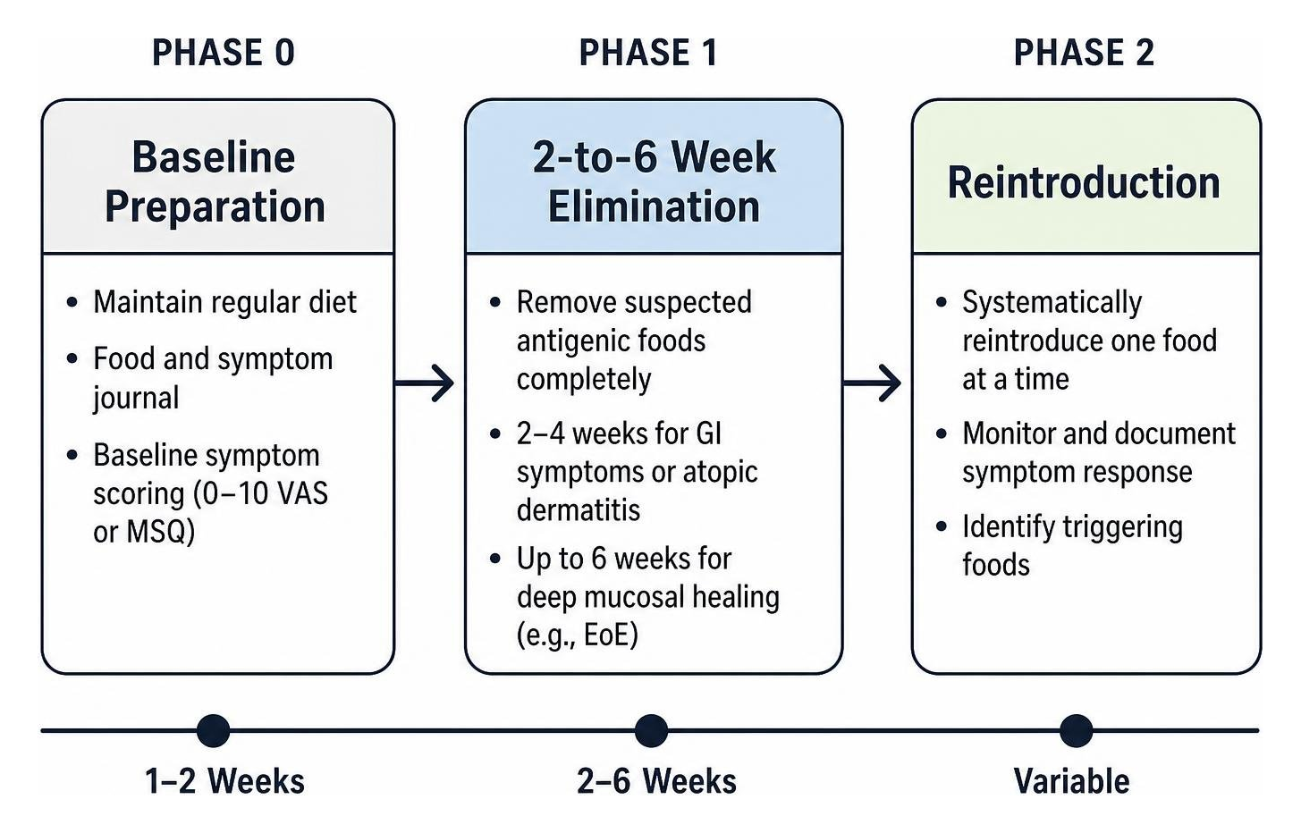
Phase 0: Baseline Characterization (1–2 Weeks): The patient maintains their regular diet while completing a detailed food and symptom journal. Objective baseline scoring (using a 0–10 Visual Analog Scale or formal Medical Symptoms Questionnaire) captures symptom frequency and intensity across cutaneous, respiratory, neurologic, and gastrointestinal domains.
Phase 1: The Elimination Window (2–6 Weeks): Suspected antigenic foods are completely removed. For functional gastrointestinal symptoms or atopic dermatitis, a 2-to-4-week window generally suffices. For conditions requiring deep mucosal healing, such as EoE, a full 6-week elimination window is required to clear residual inflammatory markers. Clinicians can review our phase 1 elimination guidelines for specific dietary onboarding workflows, or deploy a structured 21-day elimination plan for rapid clinical resets.
Maintaining Nutritional Adequacy and Safety
Eliminating major dietary groups introduces risks of nutritional deficiencies, energy deficits, and psychological distress if not actively managed. Practitioners should proactively map safe, nutrient-dense substitutions for every excluded food category:
Excluded Food Group | Potential Nutrient Gaps | Hypoallergenic Dietary Substitutions |
|---|---|---|
Dairy | Calcium, Vitamin D, Protein, Riboflavin | Fortified unsweetened coconut or hemp milk, leafy greens, canned bone-in wild salmon, blackstrap molasses |
Gluten / Wheat | B Vitamins, Dietary Fiber, Iron | Quinoa, buckwheat, wild rice, millet, certified gluten-free oats, sweet potatoes |
Eggs | Choline, Lutein, High-quality Albumin | Free-range poultry, pasture-raised beef, chia/flax seeds, pumpkin seeds |
Soy & Legumes | Folate, Magnesium, Plant Protein | Grass-fed meats, wild seafood, organic seeds (hemp, pumpkin, sunflower), collagen peptides |
Nuts & Peanuts | Essential fatty acids, Zinc, Vitamin E | Sunflower seed butter, pumpkin seeds, olive oil, avocado, chia seeds |
Clinicians must screen for absolute and relative contraindications before initiating an elimination protocol:
Active or Past Eating Disorders: Patients with anorexia nervosa, bulimia, or Avoidant/Restrictive Food Intake Disorder (ARFID) should not undergo broad elimination diets, as dietary restriction can trigger disordered eating behaviors.
Pediatric and Pregnancy Considerations: Growing children and pregnant/lactating women require strict supervision by a registered dietitian to prevent growth stunting, micronutrient deficiencies, and caloric deficits. Utilizing a nutrient-dense anti-inflammatory food list ensures patients preserve micronutrient density throughout the diagnostic window.
Step-by-Step Reintroduction: Safe Provocation and Symptom Tracking
The elimination phase only removes baseline inflammatory "noise"; the reintroduction phase provides diagnostic clarity. Systematic reintroduction transforms the elimination protocol into a controlled clinical challenge.
Foods must be reintroduced one at a time in an isolated, pure form (e.g., pure cooked egg white/yolk, plain organic cow's milk, or unflavored boiled wheat noodles), rather than complex mixed meals (e.g., baked pastries containing flour, butter, and eggs simultaneously).
Day 1 (Challenge Day - Escalating Dosing):
Morning (Dose 1): Ingest 1 teaspoon (or a small bite) of the test food on an empty stomach. Wait 2 hours while monitoring for immediate signs (itching, urticaria, rhinitis, abdominal cramping).
Midday (Dose 2): If no symptoms emerge, consume a half-portion (e.g., 1/2 cup or small serving).
Evening (Dose 3): If tolerated, consume a full normal portion with dinner.
Days 2 to 3 (Washout and Observation Window):
Discontinue the test food entirely and return strictly to the Phase 1 elimination baseline.
Monitor the patient across a 48-to-72-hour window for delayed symptom manifestations, such as altered bowel habits, migraine flares, eczema exacerbations, or joint pain.
Decision Point (Day 4):
No Symptoms: The food is marked as a tolerated non-trigger and set aside. Proceed to the next test food.
Symptoms Occur: The food is identified as a probable trigger. Record the reaction details in the clinical chart, halt the challenge immediately, and maintain the elimination baseline until all provoked symptoms have fully resolved (typically 3–5 days) before challenging the next food.
For an end-to-step operational walkthrough of testing calendars and patient tracking logs, see our online guide to elimination protocols.
Interpreting Results: When an Elimination Diet for Allergy Testing Requires an Oral Food Challenge
Interpreting reintroduction outcomes requires clinical discernment to distinguish between physiological food allergies, non-allergic intolerances, and nocebo responses.
Reaction Type | Underlying Mechanism | Typical Onset Window | Common Symptoms | Clinical Confirmation Strategy |
|---|---|---|---|---|
IgE-Mediated Allergy | Mast cell/basophil degranulation | Minutes to 2 hours | Urticaria, angioedema, wheezing, vomiting, anaphylaxis | Specific IgE, SPT, supervised medical food challenge |
Non-IgE Food Sensitivity | Cell-mediated, T-cell cytokine response | 4 to 72 hours | Joint pain, brain fog, fatigue, eczema flare, subacute GI distress | Structured elimination & multi-day washout reintroduction |
Enzymatic Intolerance | Inadequate digestive enzymes (e.g., lactase) | 30 minutes to 4 hours | Bloating, gas, osmotic diarrhea, localized cramping | Breath testing (hydrogen/methane), targeted enzyme trial |
Nocebo / Food Anxiety | Autonomic arousal, hypervigilance | Immediate to variable | Palpitations, globus sensation, subjective nausea | Blinded challenge, gradual behavioral reintroduction |
When clinical history and elimination outcomes remain ambiguous—or when distinguishing an IgE-mediated allergy from profound psychological food anxiety (nocebo effect)—the Double-Blind Placebo-Controlled Food Challenge (DBPCFC) serves as the recognized medical gold standard. Conducted in specialized clinical units equipped with emergency resuscitation protocols, the DBPCFC presents the patient with masked doses of the suspected antigen or an inert matrix (such as vehicle food powders in opaque capsules) to verify true objective reactivity without bias.
Overcoming Adherence Barriers and Tracking Clinical Outcomes
A diagnostic diet trial is only as valid as patient compliance. Minor accidental ingestions can trigger mast cell activation or digestive flares, resetting the diagnostic clock.

Common real-world adherence barriers include:
Hidden Ingredients and Cross-Contamination: Soy lecithin in chocolate, casein in processed lunch meats, wheat starch in salad dressings, and shared restaurant cooking oils can invalidate an elimination phase. Clinicians must educate patients on comprehensive ingredient label reading.
The "Mid-Trial Slump": Patient motivation often wanes around weeks 2 to 3, particularly before visible cutaneous or digestive improvements appear.
Social and Environmental Pressures: Dining outside the home poses high risks of accidental exposure. Providing patients with pre-planned dining-out strategies and meal templates minimizes accidental exposures.
Clinicians scaling functional medicine services can utilize our structured elimination diet program to deploy automated patient check-ins, symptom logging templates, and educational assets. When patients present with diffuse complaints like brain fog and chronic fatigue, applying targeted elimination strategies for systemic energy and fatigue allows practitioners to systematically correlate energy improvements with specific dietary triggers.
Frequently Asked Questions About Food Elimination Protocols
How long should the elimination phase last before testing triggers?
The standard diagnostic elimination phase ranges from 2 to 6 weeks. Gastrointestinal symptoms associated with functional gut disorders typically show meaningful improvement within 2 to 4 weeks. Chronic dermatological manifestations (such as eczema) and esophageal tissue healing in eosinophilic esophagitis generally require 6 full weeks of strict elimination. Maintaining complete dietary restriction beyond 6 weeks without initiating reintroductions provides no added diagnostic value and increases the risk of micronutrient deficiencies and microbiome alterations.
Can an elimination diet identify non-IgE food sensitivities?
Yes. An elimination diet is one of the most clinically reliable diagnostic tools for non-IgE-mediated food sensitivities. Because non-IgE reactions are driven by delayed, cell-mediated pathways (involving T-cells, localized cytokines, and eosinophils) rather than circulating IgE antibodies, blood and skin tests frequently return negative. By removing baseline antigenic exposure and systematically re-exposing the immune system, clinicians can directly observe delayed cutaneous, digestive, and neurological manifestations, including managing diet-induced anxiety.
What is the safest way to reintroduce high-risk allergens?
High-risk allergens—specifically foods associated with previous acute hives, facial angioedema, respiratory distress, or systemic anaphylaxis—must never be reintroduced at home. Reintroduction of suspected IgE-mediated allergens should occur solely under the direct supervision of a board-certified allergist in a medical setting equipped with intramuscular epinephrine, oxygen, and emergency airway support. For non-severe sensitivities, home reintroduction follows an escalating-dose schedule on Day 1, followed by a mandatory 48-to-72-hour washout observation window.
Conclusion
An elimination diet remains a cornerstone of precision clinical nutrition. While laboratory panels provide supportive data regarding sensitization and gastrointestinal inflammation, the structured process of dietary tracking, elimination, and systematic provocation delivers direct, reproducible evidence of clinical reactivity.
For licensed healthcare practitioners, executing diagnostic elimination diets with clear patient guidance, balanced nutritional substitutions, and structured tracking logs bridges the gap between complex immune mechanisms and practical patient care. To integrate these clinical tools into your practice, download our clinical Elimination Diet Food Plan to support safe, repeatable patient outcomes.
References